1. CT Scans (Computed Tomography Scans)
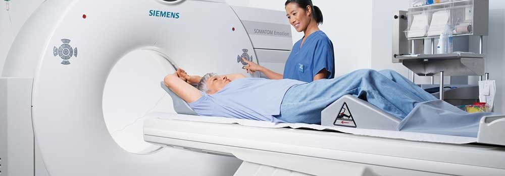
Multiple X-rays are used to deliver detailed images from inside the body as cross-sections. Bones, organs, and tumors can all be revealed quite clearly.
Major advantages of CT scans include it being a very painless process. In addition, the number of conditions which can be diagnosed as a result of these multiple rays and radiology cloud are more compared to a single X-ray. The scan is not only quick, but it also helps in figuring out if there is regrowth of a problem in a certain area inside the body.
However, higher dosage of X-rays can expose individuals to a greater risk of cancer. The dye used for contrasting is also known for causing kidney problems, and depending on what part is being scanned, the patient may need anesthesia.
2. PET (Positron-Emission Tomography)

A radioactive substance is injected/inhaled by the patient. The emitted rays are captured and processed to see images of the bones and the organs.
The number of conditions which can be identified painlessly are many when it comes to PET. Considering how the substance can cause emissions from all regions from inside the body, a problem can be identified in the very early stages, meaning that progressing towards a cure becomes a quicker step. Finally, it can also be used to determine if a given treatment is having desired results.
Gamma-rays, however, still have substantial ionization power, or might trigger allergic reactions from the sites of injection for many people. Furthermore, PET scanners have been known to cause a feeling of claustrophobia, for which further treatment becomes in order.
3. X-ray Scan

Despite being practiced less and less in the developed world, X-rays are still handy for showing dense tumors and regions inside the body coupled with radiology cloud systems they are more efficient.
While X-rays can be used efficiently and cheaply for identifying a number of physical issues inside the body such as bone fractures and tumors of some cancers, individuals (especially children) are exposed to the risk of cancer.
4. MRI (Magnetic Resonance Imaging)

MRI utilizes magnetic fields and radio waves for images of bones, cartilage, possible soft-tissue injuries, and organs from inside the body.
The most widely acclaimed advantage is the zero-degree exposure to ionizing rays, and the result is very similar to a standard CT scan in many ways. Given how it is a completely non-invasive technique, the patient does not have to suffer from any discomfort.
On the other hand, MRI requires the patient to be very still and is a lengthy process (making it difficult to get completely accurate results). In addition, the specifications for an affluent scan are lengthy as well, and can make a patient feel claustrophobic.
5. Ultrasound

Ultrasound is mostly used during pregnancy to check on the health of a baby. The high-frequency sounds produce an image of the insides of the body and can be used to monitor any growth from within. With radiology cloud systems with HTML5 DICOM viewer, ultrasound – like other imaging processes – is effective.
While this method is the most non-invasive among all, the quality of imaging and particularities such as air quality, irregularities inside the body, and being empty-stomached make it quite a demanding process in itself.
